


Japan (EN)
Select your region or country.
Development of X-ray micro-imaging system at synchrotron radiation facility
Published on March 12, 2025
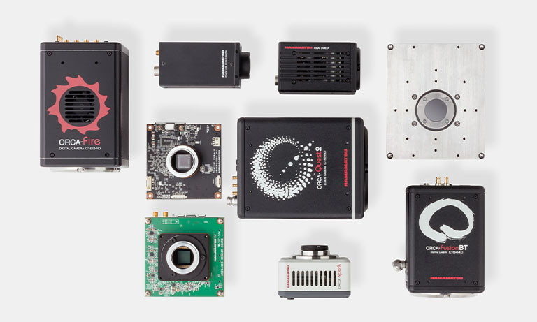
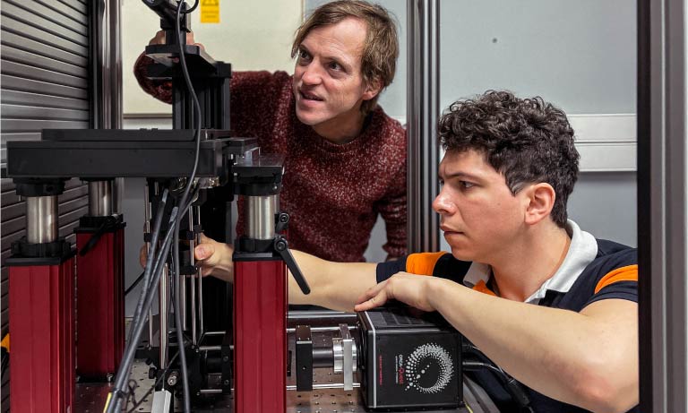
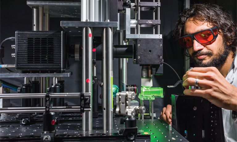
Japan Synchrotron Radiation Research Institute (JASRI) supports operating, maintaining, and utilizing the SPring-8 large synchrotron radiation facility. They also support using the SACLA X-ray free electron laser facility and the NanoTerasu 3GeV high-brilliance synchrotron radiation facility. Among them, the Microscopic and Dynamic Imaging Team, Scattering and Imaging Division, is responsible for developing X-ray imaging systems using synchrotron radiation and supporting researchers who would like to acquire X-ray micro-images. The ORCA®-Quest qCMOS® camera is used as the detector for the X-ray micro-imaging system.
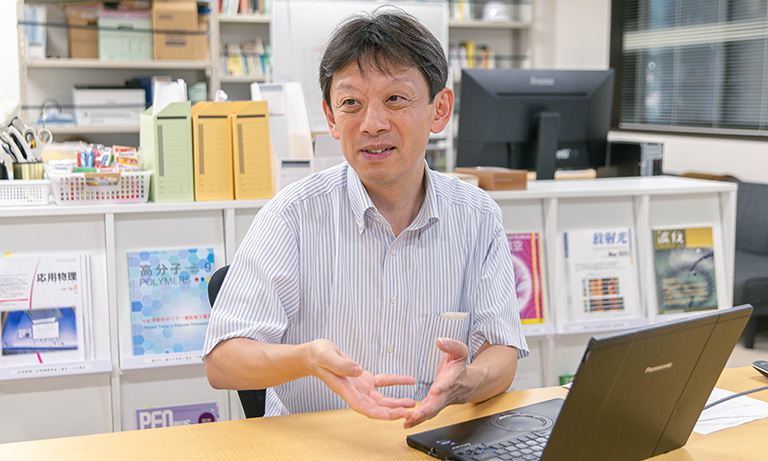
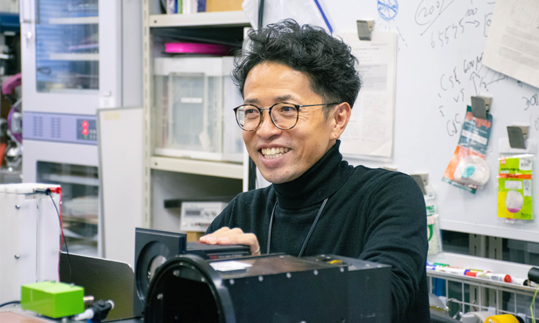
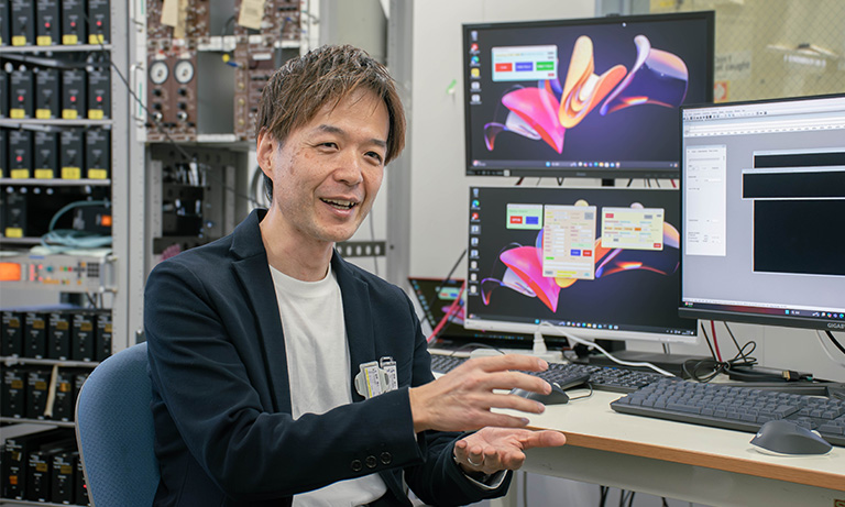
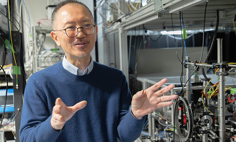
We interviewed Dr. Masato Hoshino, Senior Scientist, about the details of the work of the Microscopic and Dynamic Imaging Team, the decisive factor for introducing the ORCA-Quest, and future prospects.
The work of the Microscopic and Dynamic Imaging Team
Could you tell us about the work of the Microscopy and Dynamic Imaging Team?
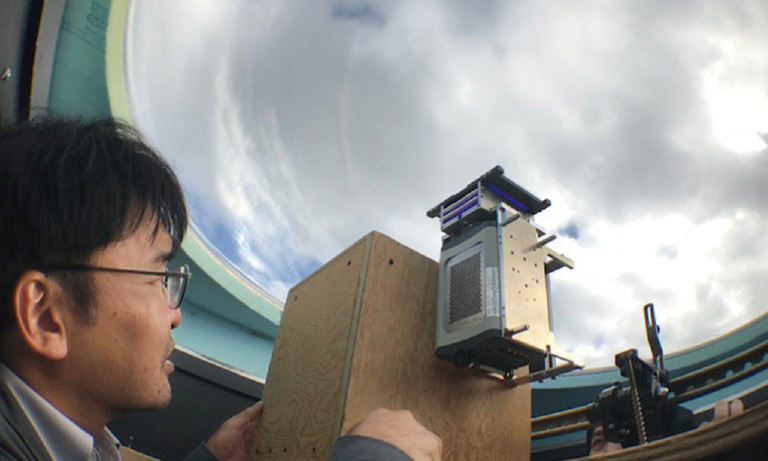
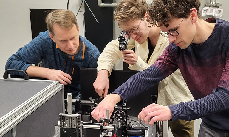
Our team is mainly engaged in developing imaging systems using X-rays and providing support for researchers who would like to use these systems. We observe a wide range of objects, including biological samples, material-based samples, excavated objects from archaeological sites, and fossils, all of which meet the demand to observe the interior of opaque objects without destroying them. At our facility, we can acquire not only 2D images such as radiographs but also 3D images using X-ray micro-tomograaphy (micro-CT).
The strength of our team is the ability to observe the interior of an object with high resolution. The large beam divergence of general X-ray sources causes the X-rays to spread out before they reach the object, resulting in a low number of photons per unit area. This results in a lower signal-to-noise ratio (S/N), making it difficult to image the fine structures inside an object with a high S/N ratio.
The X-rays generated by SPring-8 are powerful and highly directional, making it possible to irradiate narrow beams with less beam divergence. This allows for a large number of photons per unit area, to enabling high-resolution observation while maintaining a high S/N.
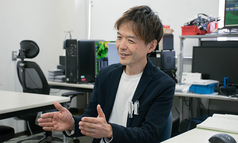
Dr. Masato Hoshino
Challenges in X-ray micro-imaging
What are the challenges of imaging with X-rays?
The important factors for X-ray micro-imaging are “field of view size,” “spatial resolution,” and “imaging speed”. Since our team supports a variety of researchers, we handle a wide range of objects, including large and small objects, objects with very fine internal structures, and objects in motion. We need to optimize the three elements according to the object, and each of these has its challenges in doing so.

When observing a large object, a wide field of view is required, but when observing an object that exceeds the camera's field of view, it is not possible to take an image with a single acquisition. Therefore, multiple images must be taken with different fields of view and then stitched together. The more images that are taken, the longer the acquisition time becomes, and the more time is required for image processing after acquisition.
To observe internal structures at high resolution, it is also important that the detector pixel size be small. However, as the pixel size becomes smaller, the amount of X-ray photons per pixel decreases. As a result, S/N decreases.
The acquisition speed greatly affects the measurement time and the observation of moving objects. For example, in X-ray micro-CT, which is used to acquire 3D images, the object is rotated and images are acquired from various angles. Sometimes thousands of images are acquired, so if the acquisition time per image is long, it will take a very long time to capture all the images. Also, when capturing images of moving objects, a slow detector frame rate can result in missing the moment we want to capture, and a long exposure time can result in blurred images.
Because there are so many objects to deal with, there are many factors that must be taken into consideration and many challenges.
Decisive factor for introducing the ORCA-Quest
Could you tell us about the decisive factor for introducing the ORCA-Quest?
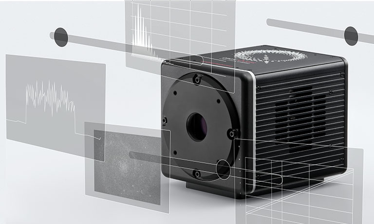
The first point is that the sensor has more pixels than conventional cameras, enabling high-definition imaging. 4096 (H) × 2304 (V) pixels, while maintaining a small pixel size of 4.6 μm × 4.6 μm, allows for high resolution and a wide field of view at the same time.
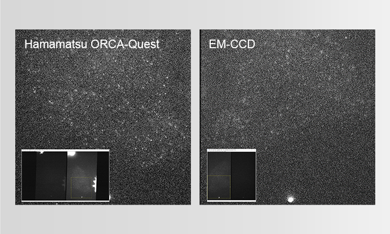
The second point is the acquisition speed. In general, increasing the frame rate of a camera reduces the number of photons per frame, resulting in a decrease in S/N. However, the ORCA-Quest not only has a high frame rate of 120 frames per second but also has high quantum efficiency and very low readout noise. As a result, it has the advantage of reducing the measurement time in X-ray micro-CT experiments and live imaging of biological specimens. In our recent experiments, we performed high-speed live imaging of the lungs of small animals.
The beamline BL20B2, where ORCA-Quest is installed, uses a bending magnet source. Bending magnet sources emit X-rays over a very wide wavelength (energy) range, so we can extract X-rays of a specific energy by a monochromator and use it. On the other hand, since the beam spreads horizontally rather than vertically, the cross-section of the beam is a horizontal shape. For this reason, a camera with a horizontal sensor shape is more compatible with nX-rays from a bending magnet source, and the sensor shape of the ORCA-Quest was fortuitously suited for this beamline.
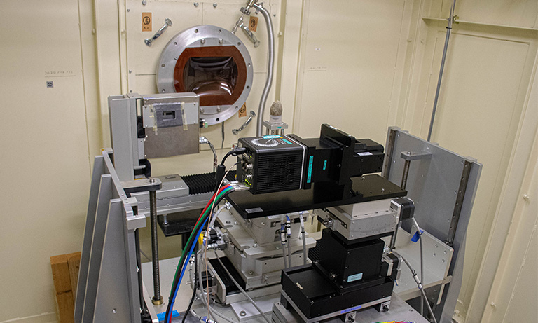
X-ray Imaging Systems
Detector: High resolution X-ray imaging system M11427, ORCA-Quest qCMOS camera C15550-20UP
Imaging example
X-ray CT image of GFRP (Glass Fiber Reinforced Plastic)

1. Transmission image of Φ8 mm GFRP rod (4096 pixels in horizontal direction, 2.68 μm/pixels)

2. Sectional image of Φ8 mm GFRP rod acquired by high-definition X-ray micro-CT (tomogram at red line in Image 1)
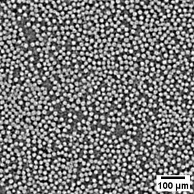
3. Enlarged image of the yellow frame area in Image 2
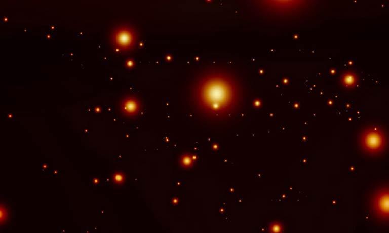
The dense concentration of glass fibers with a diameter of about 15 μm can be clearly observed.
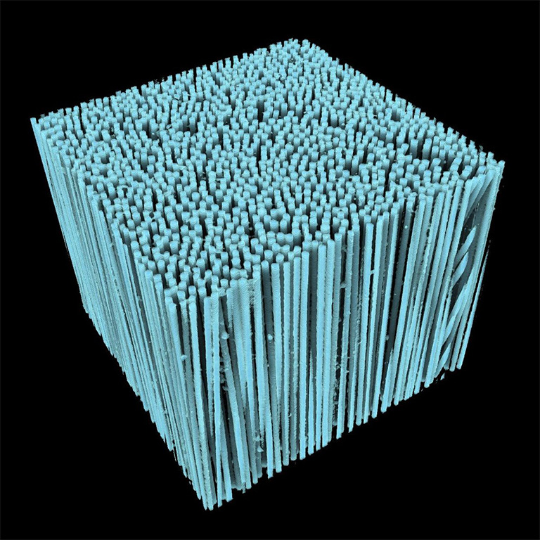
4. Glass fiber orientation displayed in 3D
The orientation of each fiber can be observed by the high-definition image.
Drishti* is used for 3D display
Camera: ORCA-Quest qCMOS camera C15550-20UP
Optics: High-resolution X-ray imaging system (M11427)
Beam line: SPring-8 BL20B2
Number of projection: 7200 images/180 degrees
Exposure time: 20 msec
Total measurement time: 3 min
Data courtesy of: SPring-8 BL20B2 beamline by Dr. Masato Hoshino, Senior Scientist in Japan Synchrotron Radiation Research Institute (JASRI)
X-ray phase-tomography image of mouse embryo
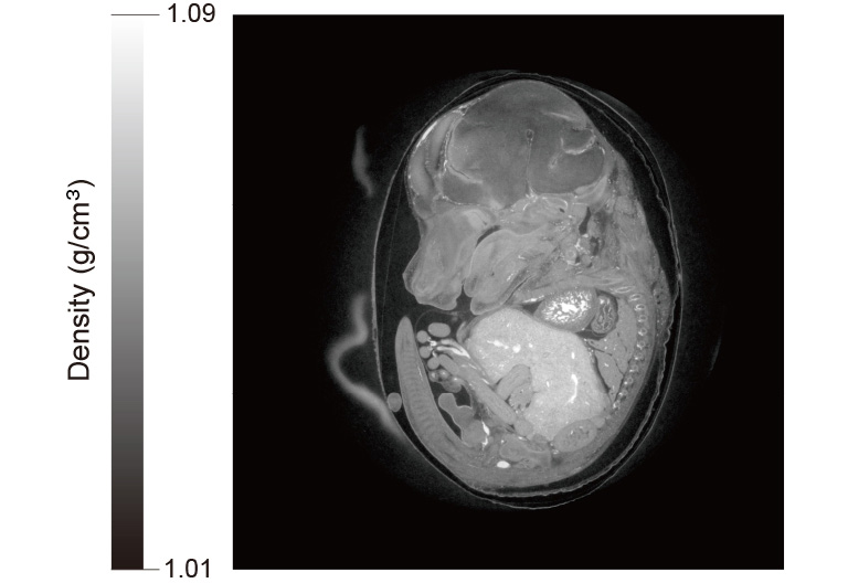
Camera: ORCA-Quest qCMOS camera C15550-20UP
Optics: High-resolution X-ray imaging system (M11427)
Beam line: SPring-8 BL20B2
Exposure time: 15 msec
Total measurement time: 6.5 min
Data courtesy of: SPring-8 BL20B2 beamline by Dr. Masato Hoshino, Senior Scientist in Japan Synchrotron Radiation Research Institute (JASRI)
Future prospects
Could you tell us about your prospects for future systems development?
Currently, I am mainly focusing on the development of X-ray micro imaging systems for multiscale measurement. Multiscale measurement means taking images at multiple spatial scales with different pixel sizes and field of views. Some researchers who use our facility want to see as much detail as possible inside large objects, such as excavations from archaeological sites or fossils. The maximum beam available at BL20B2 is about 150 mm (H) × 20 mm (V), which has the potential to observe larger objects. When acquiring X-ray images, we use a method where the X-rays are converted to visible light by a scintillator and then focused onto the sensor using a lens system. However, when balancing the field of view and spatial resolution, we have to prioritize one over the other in a single shot.
In multi scale measurement, we can observe more efficiently by first acquiring the entire object with a camera that has a large field of view, and then zooming in on the areas that require more detailed observation using the ORCA-Quest. When changing the camera, it is necessary to finely adjust the synchronization with peripheral devices such as the rotation stage on which the object is placed, as well as other settings like beam size. Therefore, we will continue to conduct verification to ensure optimal imaging under various conditions.
Of course, while maintaining the current pixel size and sensitivity, it would be ideal to use a camera with more pixels and a larger sensor size, allowing for observation with a larger field of view. Therefore, we look forward to future camera developments from Hamamatsu Photonics.
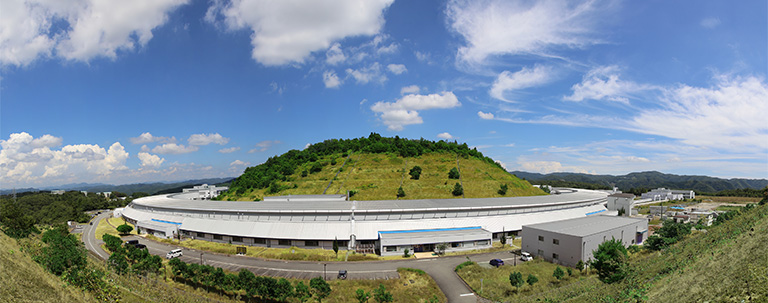
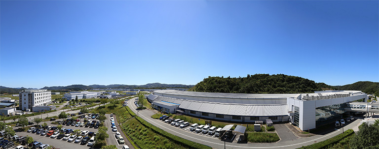
About Japan Synchrotron Radiation Research Institute (JASRI)
Japan Synchrotron Radiation Research Institute (JASRI) supports operating, maintaining, and utilizing the SPring-8 large synchrotron radiation facility. They also support using the SACLA X-ray free electron laser facility and the NanoTerasu 3GeV high-brilliance synchrotron radiation facility.
These facilities are open to a wide range of domestic and international users. They are operated by a large number of highly specialized staff to ensure smooth, high level usage of the world's most advanced synchrotron radiation facilities.
Researcher profile

Masato Hoshino
Microscopic and Dynamic Imaging Team, Scattering and Imaging Division, Japan Synchrotron Radiation Research Institute (JASRI) : Senior Scientist
Mar. 2008
Applied Physics, Graduate School of Pure and Applied Sciences, University of Tsukuba, Ph.D. (Doctor of Engineering)
Apr. 2008
Post-doctoral researcher, Research and Utilization Division, Japan Synchrotron Radiation Research Institute (JASRI)
Apr. 2009
Research scientist, Research and Utilization Division, Japan Synchrotron Radiation Research Institute (JASRI)
Apr. 2018
Senior Scientist, Research and Utilization Division, Japan Synchrotron Radiation Research Institute (JASRI)
Apr. 2021
Senior Scientist, Microscopic and Dynamic Imaging Team, Scattering and Imaging Division, Japan Synchrotron Radiation Research Institute (JASRI)
*The content presented on this page is based on an interview conducted in December 2024.
Related product

The ORCA-Quest 2 is a new qCMOS® camera, the successor to the ORCA-Quest with further advances such as faster readout speeds in extremely low-noise scan mode and increased sensitivity in the ultraviolet region.
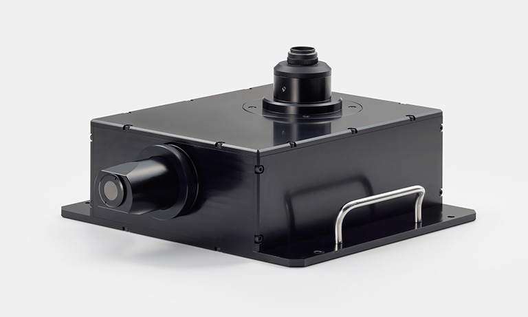
The high resolution X-ray imaging system is designed for the application of X-ray imaging in synchrotron radiation facilities. Real-time X-ray phenomena can be imaged by combining an imaging unit that uses a phosphor to visualize an incident X-ray beam, and Hamamatsu’s digital camera.
Other case studies
- Confirmation
-
It looks like you're in the . If this is not your location, please select the correct region or country below.
You're headed to Hamamatsu Photonics website for JP (English). If you want to view an other country's site, the optimized information will be provided by selecting options below.
In order to use this website comfortably, we use cookies. For cookie details please see our cookie policy.
- Cookie Policy
-
This website or its third-party tools use cookies, which are necessary to its functioning and required to achieve the purposes illustrated in this cookie policy. By closing the cookie warning banner, scrolling the page, clicking a link or continuing to browse otherwise, you agree to the use of cookies.
Hamamatsu uses cookies in order to enhance your experience on our website and ensure that our website functions.
You can visit this page at any time to learn more about cookies, get the most up to date information on how we use cookies and manage your cookie settings. We will not use cookies for any purpose other than the ones stated, but please note that we reserve the right to update our cookies.
1. What are cookies?
For modern websites to work according to visitor’s expectations, they need to collect certain basic information about visitors. To do this, a site will create small text files which are placed on visitor’s devices (computer or mobile) - these files are known as cookies when you access a website. Cookies are used in order to make websites function and work efficiently. Cookies are uniquely assigned to each visitor and can only be read by a web server in the domain that issued the cookie to the visitor. Cookies cannot be used to run programs or deliver viruses to a visitor’s device.
Cookies do various jobs which make the visitor’s experience of the internet much smoother and more interactive. For instance, cookies are used to remember the visitor’s preferences on sites they visit often, to remember language preference and to help navigate between pages more efficiently. Much, though not all, of the data collected is anonymous, though some of it is designed to detect browsing patterns and approximate geographical location to improve the visitor experience.
Certain type of cookies may require the data subject’s consent before storing them on the computer.
2. What are the different types of cookies?
This website uses two types of cookies:
- First party cookies. For our website, the first party cookies are controlled and maintained by Hamamatsu. No other parties have access to these cookies.
- Third party cookies. These cookies are implemented by organizations outside Hamamatsu. We do not have access to the data in these cookies, but we use these cookies to improve the overall website experience.
3. How do we use cookies?
This website uses cookies for following purposes:
- Certain cookies are necessary for our website to function. These are strictly necessary cookies and are required to enable website access, support navigation or provide relevant content. These cookies direct you to the correct region or country, and support security and ecommerce. Strictly necessary cookies also enforce your privacy preferences. Without these strictly necessary cookies, much of our website will not function.
- Analytics cookies are used to track website usage. This data enables us to improve our website usability, performance and website administration. In our analytics cookies, we do not store any personal identifying information.
- Functionality cookies. These are used to recognize you when you return to our website. This enables us to personalize our content for you, greet you by name and remember your preferences (for example, your choice of language or region).
- These cookies record your visit to our website, the pages you have visited and the links you have followed. We will use this information to make our website and the advertising displayed on it more relevant to your interests. We may also share this information with third parties for this purpose.
Cookies help us help you. Through the use of cookies, we learn what is important to our visitors and we develop and enhance website content and functionality to support your experience. Much of our website can be accessed if cookies are disabled, however certain website functions may not work. And, we believe your current and future visits will be enhanced if cookies are enabled.
4. Which cookies do we use?
There are two ways to manage cookie preferences.
- You can set your cookie preferences on your device or in your browser.
- You can set your cookie preferences at the website level.
If you don’t want to receive cookies, you can modify your browser so that it notifies you when cookies are sent to it or you can refuse cookies altogether. You can also delete cookies that have already been set.
If you wish to restrict or block web browser cookies which are set on your device then you can do this through your browser settings; the Help function within your browser should tell you how. Alternatively, you may wish to visit www.aboutcookies.org, which contains comprehensive information on how to do this on a wide variety of desktop browsers.
5. What are Internet tags and how do we use them with cookies?
Occasionally, we may use internet tags (also known as action tags, single-pixel GIFs, clear GIFs, invisible GIFs and 1-by-1 GIFs) at this site and may deploy these tags/cookies through a third-party advertising partner or a web analytical service partner which may be located and store the respective information (including your IP-address) in a foreign country. These tags/cookies are placed on both online advertisements that bring users to this site and on different pages of this site. We use this technology to measure the visitors' responses to our sites and the effectiveness of our advertising campaigns (including how many times a page is opened and which information is consulted) as well as to evaluate your use of this website. The third-party partner or the web analytical service partner may be able to collect data about visitors to our and other sites because of these internet tags/cookies, may compose reports regarding the website’s activity for us and may provide further services which are related to the use of the website and the internet. They may provide such information to other parties if there is a legal requirement that they do so, or if they hire the other parties to process information on their behalf.
If you would like more information about web tags and cookies associated with on-line advertising or to opt-out of third-party collection of this information, please visit the Network Advertising Initiative website http://www.networkadvertising.org.
6. Analytics and Advertisement Cookies
We use third-party cookies (such as Google Analytics) to track visitors on our website, to get reports about how visitors use the website and to inform, optimize and serve ads based on someone's past visits to our website.
You may opt-out of Google Analytics cookies by the websites provided by Google:
https://tools.google.com/dlpage/gaoptout?hl=en
As provided in this Privacy Policy (Article 5), you can learn more about opt-out cookies by the website provided by Network Advertising Initiative:
http://www.networkadvertising.org
We inform you that in such case you will not be able to wholly use all functions of our website.
Close