


Japan (EN)
Select your region or country.
Development of heavy ion imaging technology for application in hadron radiotherapy
Published on February 24, 2025
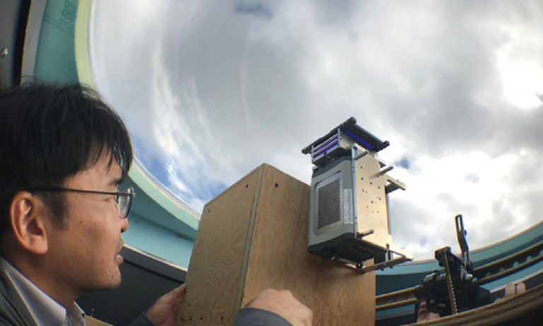
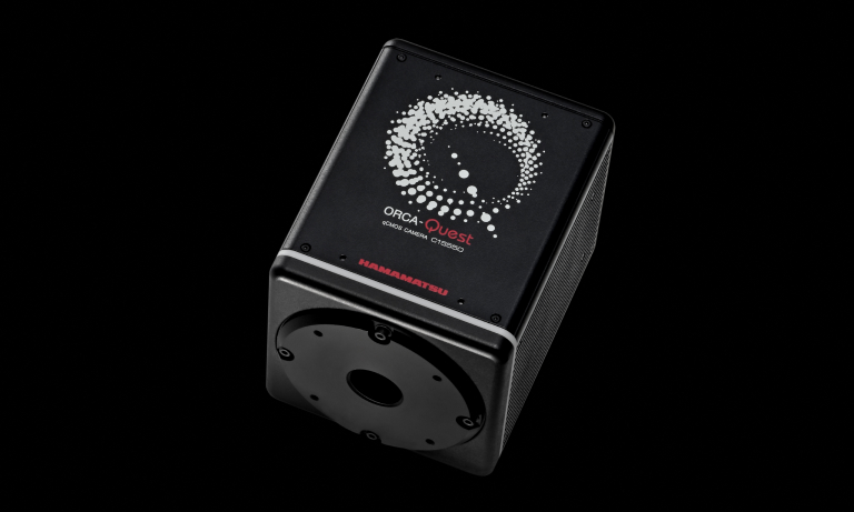
The Advanced Beam Measurement Group, Research Institute for Measurement and Analytical Instrumentation, National Metrology Institute of Japan (NMIJ), National Institute of Advanced Industrial Science and Technology (AIST) conducts research to develop advanced measurement and analysis technologies using quantum beams such as X-rays, positrons, and neutrons to contribute to materials development and the realization of a safe and secure society. One of the research themes in this laboratory is the development of alpha-ray imaging technology for advanced heavy ion radiotherapy, in which our ORCA®-Quest qCMOS® camera is used as a detector.
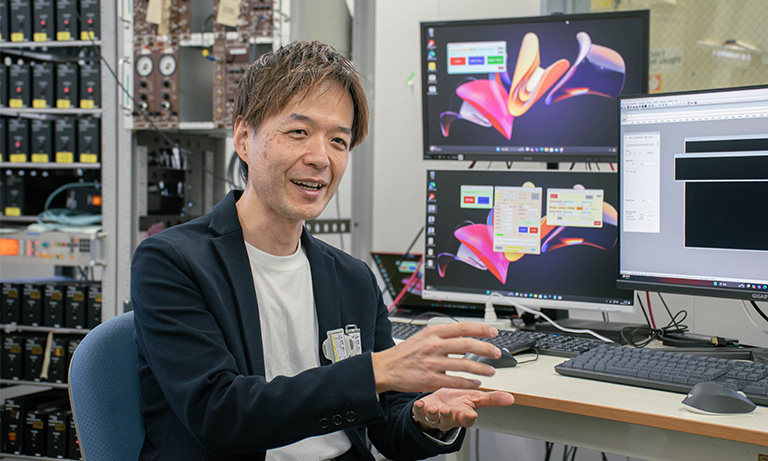
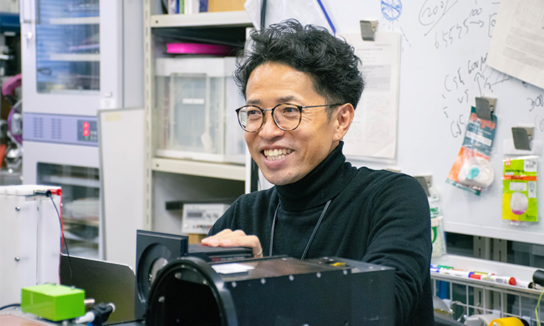
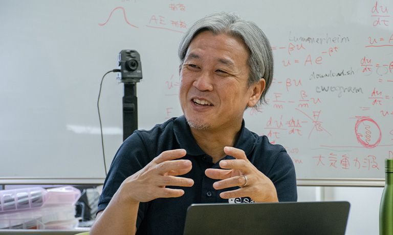
We interviewed Dr. Takeshi Fujiwara, Group Leader of the research group, about the details of the research, the results obtained by using ORCA-Quest, and future research prospects.
Current research
Could you tell us about your research?
Our group is developing dose imaging technology for hadron therapy to improve the accuracy of dose distribution in hadron radiotherapy.
In recent years, hadron radiotherapy using carbon ions has gained attention as an effective treatment method among radiation therapies. Currently, X-rays are primarily used as the radiation source in radiation therapy. However, X-rays have the characteristic of delivering the highest dose at the body's surface, with the dose decreasing as they penetrate deeper into the body where the cancerous tissue is located. This results in the cancerous tissue and the normal tissues at the body's surface being exposed to radiation.
On the other hand, heavy ion beams such as alpha-ray and carbon ions have a characteristic called the Bragg peak, where the dose is low at the body's surface and increases as it approaches the target tissue. This allows for selective irradiation of the cancerous cells without affecting the normal cells at the body's surface. Consequently, various institutions are currently developing carbon ion irradiation technology. To irradiate these heavy ion beams at the target location, it is necessary to measure with high precision the intensity and location of the irradiation and use the results to adjust the source intensity and irradiation location.
However, there are various challenges in the current technology for high precision imaging and measurement of heavy ion beams, and it has not yet been established. Our laboratory is working on developing new imaging technologies to address these challenges.
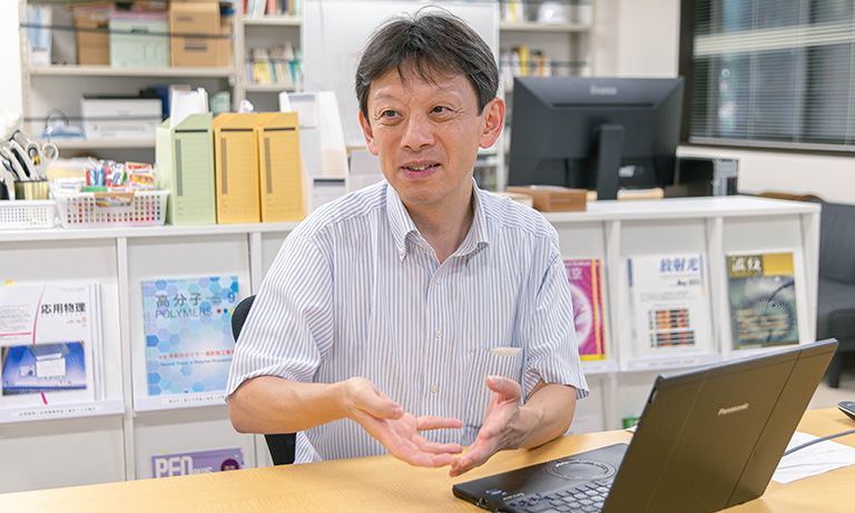
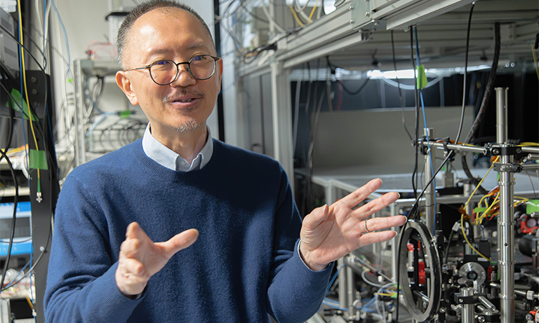
Dr. Takeshi Fujiwara
Challenges in imaging and measuring heavy particle beams

Conceptual diagram of Glass GEM
Data provided by: Dr. Takeshi Fujiwara, The Advanced Beam Measurement Group, Research Institute for Measurement and Analytical Instrumentation, National Metrology Institute of Japan (NMIJ), National Institute of Advanced Industrial Science and Technology (AIST)
What are the challenges in imaging measurement of heavy particle beams?
Currently, ionization chambers are the primary detectors used for heavy ion beam measurements. However, as point detectors, ionization chambers inherently lack spatial resolution and cannot be used for measuring 2D dose distributions. While array-type ionization chambers have recently been developed, they have limitations: their spatial resolution is only about 5 mm, and they require numerous circuits for multi-channel readout, which not only creates implementation challenges but also increases costs significantly.
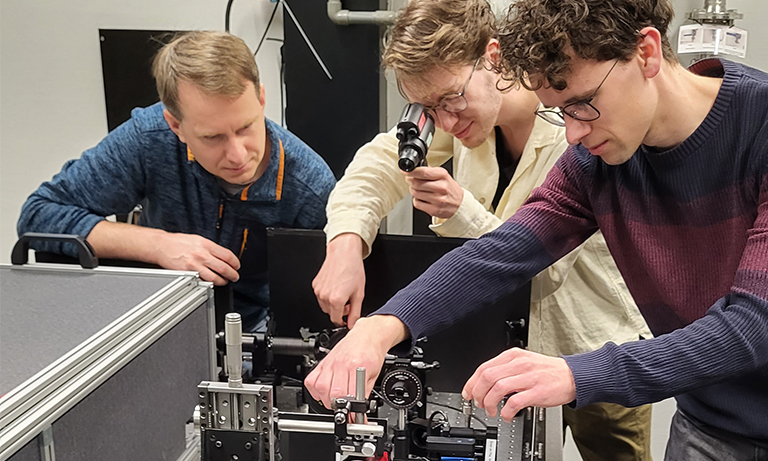
To address these limitations, our laboratory is developing a new detector system that combines GEM (Gas Electron Multiplier) technology with high-sensitivity cameras. While GEM was originally developed as a 2D radiation detector that amplifies electrons using gas, our group's Glass GEM achieves higher amplification rates compared to conventional GEMs. Furthermore, by combining it with an Ar/CF4 fluorescent gas mixture, it performs high-sensitivity and high-resolution imaging of heavy ion beams. Using this Glass GEM with Hamamatsu Photonics' ORCA-Quest qCMOS camera, we have achieved heavy ion beam imaging with exceptionally high signal-to-noise ratios and high spatial resolution. Specifically, while detectors using ionization chamber arrays have a spatial resolution of about 5 mm, our detector system using the Glass GEM and camera achieves a spatial resolution of approximately 1 mm.
Results obtained using ORCA-Quest
What results have you seen from using ORCA-Quest?
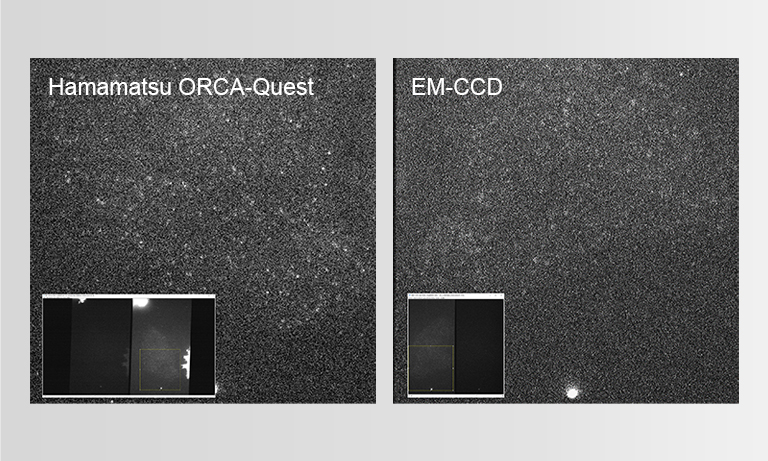
Before using the ORCA-Quest, we used Hamamatsu Photonics' ORCA-Flash4.0 V3. While this camera, when combined with GEM, could image alpha rays which is a type of particle radiation, the images lacked contrast due to insufficient sensitivity and noise (Imaging example 1).
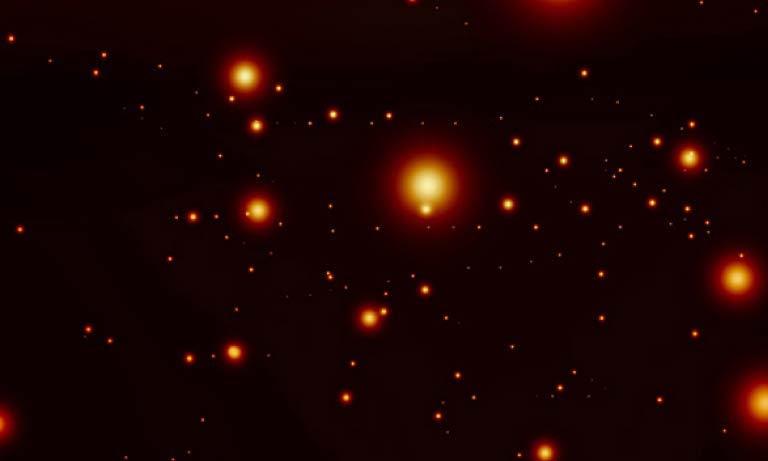
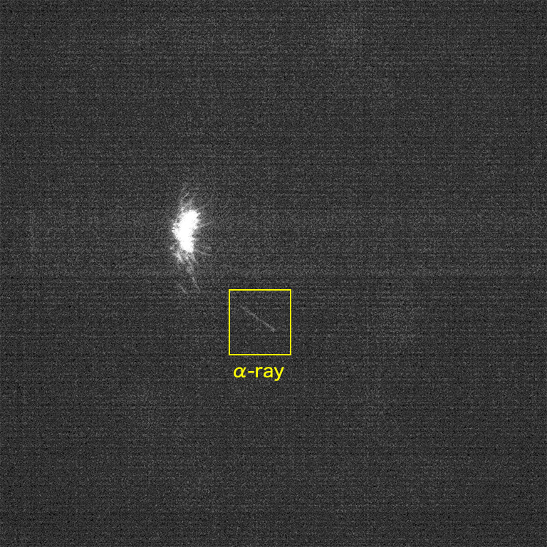
When we switched to the ORCA-Quest camera, we observed a dramatic improvement in the signal-to-noise ratio compared to the ORCA-Flash4.0 V3. Imaging example 2 shows alpha ray imaging using the ORCA-Quest, where we can not only visualize individual alpha rays as they arrive but also capture subtle variations in dose distribution with high precision. The images clearly show the highest dose concentration at the track endpoints due to the Bragg peak effect. Conventional alpha ray detection methods could only produce integrated images showing faint glowing regions, making it difficult to distinguish between the presence of zero or one alpha particle. Therefore, our system's ability to achieve this level of detection precision while simultaneously performing imaging represents a groundbreaking advancement in the field.
In the future, when this detector is implemented in medical devices and treatment technologies, it will need to meet extremely high precision requirements as it will directly impact human lives. We believe the superior performance of our system will prove invaluable in meeting these demanding requirements.
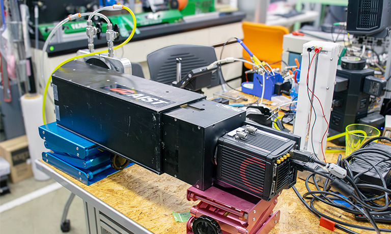
Glass GEM and camera combined detector
Imaging example
1. Visualized image of alpha radiation using ORCA-Flash4.0 V3.
Data courtesy of: Dr. Takeshi Fujiwara, The Advanced Beam Measurement Group, Research Institute for Measurement and Analytical Instrumentation, National Metrology Institute of Japan (NMIJ), National Institute of Advanced Industrial Science and Technology (AIST).
2. Visualized image of alpha radiation using ORCA-Quest.
Scan mode: Standard scan
Exposure time: 10 ms
Interval: 14.12 ms
Number of pixels used: 1024 (H) × 576 (V)
Data courtesy of: Dr. Takeshi Fujiwara, The Advanced Beam Measurement Group, Research Institute for Measurement and Analytical Instrumentation, National Metrology Institute of Japan (NMIJ), National Institute of Advanced Industrial Science and Technology (AIST).
Prospects for research
Could you tell us about your future research prospects?
Future prospects for heavy ion radiotherapy include the following. We hope to contribute to the advancement of these research and development efforts by using the detector developed by our laboratory.
- 3D heavy ion irradiation using spot scanning
- Development of treatment techniques with fewer side effects using FLASH irradiation
Regarding spot scanning: In conventional heavy ion therapy, the beam is collimated according to the size of the tumor and irradiated. While heavy ion beams offer the advantage of minimal impact on the body's surface tissue, they still pose the challenge of exposing healthy cells around the tumor. To address this issue, a new approach has been proposed: shaping the heavy ion beam into a thin beam called a 'pencil beam' and dynamically scanning it in the X-Y direction to deliver localized treatment. Moreover, by varying the dose, we can adjust the irradiation depth, enabling 3D beam delivery to the tumor.
To verify whether the pencil beam is irradiated at the targeted position in 3D, a detector developed by our laboratory that combines a Glass GEM and a camera is used. This detector can simultaneously and dynamically detect the 2D distribution of the heavy particle beam and the dose intensity, thus accurately determining the 3D dose distribution. We expect that this detector will further develop the spot scanning technique in the future.
Regarding FLASH irradiation: Currently, researchers are developing a treatment method with reduced side effects using "FLASH irradiation." This technique involves delivering an extremely high dose – hundreds of times higher than conventional doses – to the tumor in a very brief moment. Experimental results have shown that compared to prolonged exposure at lower doses, FLASH irradiation maintains therapeutic effectiveness while reducing adverse effects on the human body. While FLASH irradiation requires delivering high doses in very short timeframes, achieving this requires operating synchrotron accelerators in somewhat unconventional ways. Consequently, there remains uncertainty about whether the beam can be delivered at the intended intensity and to the precise target location. We believe our detector can also be valuable in monitoring these parameters.

Researcher profile

Dr. Takeshi Fujiwara
Advanced Beam Measurement Group, Research Institute for Measurement and Analytical Instrumentation, National Metrology Institute of Japan (NMIJ), National Institute of Advanced Industrial Science and Technology (AIST): Group Leader
Mar. 2009
Department of Nuclear Engineering and Management, Graduate School of Engineering, The University of Tokyo, Ph.D.
Apr. 2009
Project Researcher, Department of Nuclear Engineering and Management, Graduate School of Engineering, The University of Tokyo
Apr. 2010
Assistant Professor, Department of Nuclear Professional School, Graduate School of Engineering, The University of Tokyo
Apr. 2015
Research Scientist, Research Institute for Measurement and Analytical Instrumentation, National Institute of Advanced Industrial Science and Technology (AIST)
Oct. 2019
Senior Research Scientist, Research Institute for Measurement and Analytical Instrumentation, National Institute of Advanced Industrial Science and Technology (AIST)
Apr. 2023
Principal Research Scientist, Research Institute for Measurement and Analytical Instrumentation, National Institute of Advanced Industrial Science and Technology (AIST)
Oct. 2023
Principal Research Manager, Research Strategy Planning Division, National Institute of Advanced Industrial Science and Technology (AIST)
Oct. 2024
Group Leader, Advanced Beam Measurement Group, Research Institute for Measurement and Analytical Instrumentation, National Metrology Institute of Japan (NMIJ), National Institute of Advanced Industrial Science and Technology (AIST)
*The content presented on this page is based on an interview conducted in December 2024.
Related product
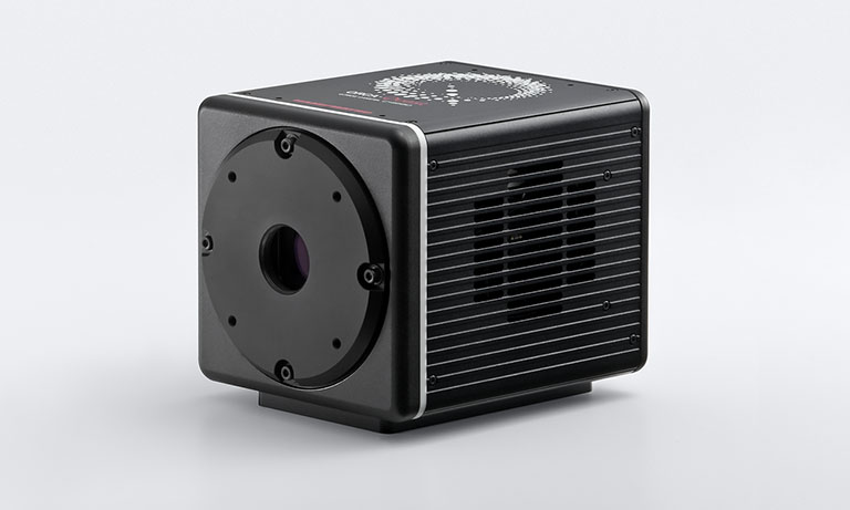
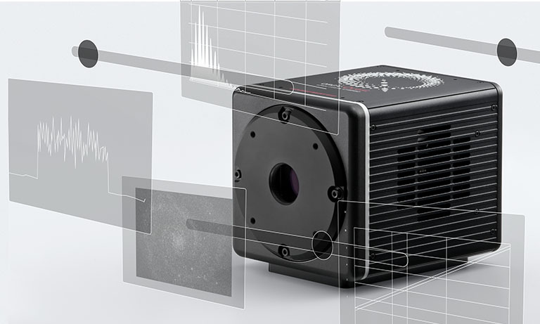
The ORCA-Quest 2 is a new qCMOS® camera, the successor to the ORCA-Quest with further advances such as faster readout speeds in extremely low-noise scan mode and increased sensitivity in the ultraviolet region.
Other case studies
- Confirmation
-
It looks like you're in the . If this is not your location, please select the correct region or country below.
You're headed to Hamamatsu Photonics website for JP (English). If you want to view an other country's site, the optimized information will be provided by selecting options below.
In order to use this website comfortably, we use cookies. For cookie details please see our cookie policy.
- Cookie Policy
-
This website or its third-party tools use cookies, which are necessary to its functioning and required to achieve the purposes illustrated in this cookie policy. By closing the cookie warning banner, scrolling the page, clicking a link or continuing to browse otherwise, you agree to the use of cookies.
Hamamatsu uses cookies in order to enhance your experience on our website and ensure that our website functions.
You can visit this page at any time to learn more about cookies, get the most up to date information on how we use cookies and manage your cookie settings. We will not use cookies for any purpose other than the ones stated, but please note that we reserve the right to update our cookies.
1. What are cookies?
For modern websites to work according to visitor’s expectations, they need to collect certain basic information about visitors. To do this, a site will create small text files which are placed on visitor’s devices (computer or mobile) - these files are known as cookies when you access a website. Cookies are used in order to make websites function and work efficiently. Cookies are uniquely assigned to each visitor and can only be read by a web server in the domain that issued the cookie to the visitor. Cookies cannot be used to run programs or deliver viruses to a visitor’s device.
Cookies do various jobs which make the visitor’s experience of the internet much smoother and more interactive. For instance, cookies are used to remember the visitor’s preferences on sites they visit often, to remember language preference and to help navigate between pages more efficiently. Much, though not all, of the data collected is anonymous, though some of it is designed to detect browsing patterns and approximate geographical location to improve the visitor experience.
Certain type of cookies may require the data subject’s consent before storing them on the computer.
2. What are the different types of cookies?
This website uses two types of cookies:
- First party cookies. For our website, the first party cookies are controlled and maintained by Hamamatsu. No other parties have access to these cookies.
- Third party cookies. These cookies are implemented by organizations outside Hamamatsu. We do not have access to the data in these cookies, but we use these cookies to improve the overall website experience.
3. How do we use cookies?
This website uses cookies for following purposes:
- Certain cookies are necessary for our website to function. These are strictly necessary cookies and are required to enable website access, support navigation or provide relevant content. These cookies direct you to the correct region or country, and support security and ecommerce. Strictly necessary cookies also enforce your privacy preferences. Without these strictly necessary cookies, much of our website will not function.
- Analytics cookies are used to track website usage. This data enables us to improve our website usability, performance and website administration. In our analytics cookies, we do not store any personal identifying information.
- Functionality cookies. These are used to recognize you when you return to our website. This enables us to personalize our content for you, greet you by name and remember your preferences (for example, your choice of language or region).
- These cookies record your visit to our website, the pages you have visited and the links you have followed. We will use this information to make our website and the advertising displayed on it more relevant to your interests. We may also share this information with third parties for this purpose.
Cookies help us help you. Through the use of cookies, we learn what is important to our visitors and we develop and enhance website content and functionality to support your experience. Much of our website can be accessed if cookies are disabled, however certain website functions may not work. And, we believe your current and future visits will be enhanced if cookies are enabled.
4. Which cookies do we use?
There are two ways to manage cookie preferences.
- You can set your cookie preferences on your device or in your browser.
- You can set your cookie preferences at the website level.
If you don’t want to receive cookies, you can modify your browser so that it notifies you when cookies are sent to it or you can refuse cookies altogether. You can also delete cookies that have already been set.
If you wish to restrict or block web browser cookies which are set on your device then you can do this through your browser settings; the Help function within your browser should tell you how. Alternatively, you may wish to visit www.aboutcookies.org, which contains comprehensive information on how to do this on a wide variety of desktop browsers.
5. What are Internet tags and how do we use them with cookies?
Occasionally, we may use internet tags (also known as action tags, single-pixel GIFs, clear GIFs, invisible GIFs and 1-by-1 GIFs) at this site and may deploy these tags/cookies through a third-party advertising partner or a web analytical service partner which may be located and store the respective information (including your IP-address) in a foreign country. These tags/cookies are placed on both online advertisements that bring users to this site and on different pages of this site. We use this technology to measure the visitors' responses to our sites and the effectiveness of our advertising campaigns (including how many times a page is opened and which information is consulted) as well as to evaluate your use of this website. The third-party partner or the web analytical service partner may be able to collect data about visitors to our and other sites because of these internet tags/cookies, may compose reports regarding the website’s activity for us and may provide further services which are related to the use of the website and the internet. They may provide such information to other parties if there is a legal requirement that they do so, or if they hire the other parties to process information on their behalf.
If you would like more information about web tags and cookies associated with on-line advertising or to opt-out of third-party collection of this information, please visit the Network Advertising Initiative website http://www.networkadvertising.org.
6. Analytics and Advertisement Cookies
We use third-party cookies (such as Google Analytics) to track visitors on our website, to get reports about how visitors use the website and to inform, optimize and serve ads based on someone's past visits to our website.
You may opt-out of Google Analytics cookies by the websites provided by Google:
https://tools.google.com/dlpage/gaoptout?hl=en
As provided in this Privacy Policy (Article 5), you can learn more about opt-out cookies by the website provided by Network Advertising Initiative:
http://www.networkadvertising.org
We inform you that in such case you will not be able to wholly use all functions of our website.
Close