


United States (EN)
Select your region or country.
How much resolution? How wide a field-of-view?
THIS GUIDE IS FOR:
Biologists that use, or are interested in using, microscope cameras and don’t speak engineering
THIS GUIDE OFFERS:
Clarity on the relevance of camera specs to biological experimentation
SECTIONS:
LISTED IN SPECIFICATIONS AS:
| Hamamatsu listing | Synonyms used by other vendors |
|---|---|
| Effective pixels | Active pixels, Resolution |
| Cell size | Pixel size |
| Effective area | Image area, Active area, Sensor/diagonal |
Field-of-view
Reading a camera specifications table can involve not only having to translate from engineering into biology, but from the language of one camera manufacturer to another. This is particularly true of the terms that go into describing how much resolution is achievable.
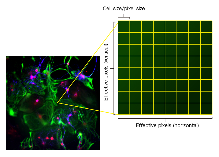
Let’s start with the terms effective pixels, active pixels, and resolution. These all refer to the number of pixels that go into capturing an image—the square or rectangular grid that you can lay over your image, where each pixel reports on photons captured over a specific area (see figure below).
The area of the image that each pixel covers depends on the size of the pixel and is listed in the specifications table as cell size or pixel size.
Multiplying the cell/pixel size by the number of pixels converts the field-of-view captured in a single image from pixels to millimeters, and is listed as effective area, image area, active area, or sensor format/diagonal.
Why are the number of pixels described as effective pixels or active pixels instead of simply pixels? The words effective and active are used to remove any ambiguity on exactly how many pixels are actually used to capture an image—for some cameras not all the pixels on the sensor contribute to the image, but rather are used for transferring or holding photoelectrons or voltage.
Resolution
How does the pixel size spec translate into camera resolution?
For starters, it’s important to bear in mind that the optical system limits resolution—if the microscope can’t resolve the image then the camera will not be able to see it either. So we begin by looking at microscope resolution and calculating the size of the projected image of the object to be resolved. And to do this, we need to first define what we mean by resolution.
How is resolution defined?
In 1873, the physicist Ernst Abbe first defined the diffraction limit of an objective as l/2NA. Lord Rayleigh later refined this equation for microscopes to be 0.61 × l/NA—the separation necessary to distinguish two Airy patterns as separate objects.
From Rayleigh’s equation we can calculate the size of the smallest item the objective can resolve, and when this number is multiplied by the magnification of the objective (assuming no additional intermediate optics) we can calculate how large the item will appear to be when it is projected onto the camera’s sensor.
What size pixels do I need to see this level of resolution?
Now that we have determined the resolution of the optical system and the projected size of objects, we can determine the maximum acceptable pixel size needed to meet this resolution. The question becomes one of sampling—given the need to fulfill the Nyquist criteria, which provides a theoretical framework for the minimum number of pixels needed to accurately define an object, we need to ensure that the object of interest is projected on to at least two pixels on the camera sensor (ideally more, 2.5 to 3 is recommended for high resolution microscopy.)
As an example, we’ll imagine trying to resolve peroxisomes from rat liver parenchymal cells that are roughly 0.7 μm in diameter. To resolve a 0.7 μm object, we’d need an objective of similar or higher resolution. From Table 1, the minimum magnification needed would be the objective with10× magnification and an NA of 0.45 (of course, if a wide field-of-view is not needed, most microscopists would oversample and use a higher magnification to achieve a more visually pleasing and informative image). At this magnification, the projected image would be 7.5 μm, so we’d need a camera with a pixel size of 7.5 μm / 2 = 3.7 μm or smaller.
| Table 1. | ||||
|---|---|---|---|---|
| Objective | Numerical Aperture (NA) | Resolution Limit*(Rayleigh’s criterion) (μm) | Projected Size (μm) | Maximum Acceptable Pixel Size (μm) |
| 4 | 0.20 | 1.7 | 6.7 | 3.4 |
| 10 | 0.30 | 1.1 | 11.2 | 5.6 |
| 10 | 0.45 | 0.7 | 7.5 | 3.7 |
| 20 | 0.50 | 0.7 | 13.4 | 6.7 |
| 20 | 0.75 | 0.4 | 8.9 | 4.5 |
| 40 | 0.75 | 0.4 | 17.9 | 8.9 |
| 40 | 1.30 | 0.3 | 10.3 | 5.2 |
| 60 | 1.40 | 0.2 | 14.4 | 7.2 |
| 100 | 1.30 | 0.3 | 25.8 | 12.9 |
| 100 | 1.40 | 0.2 | 24.0 | 12.0 |
| *Assume 550 nm light | ||||
BOTTOM LINE:
- The field-of-view that a camera is able to deliver is reflected in the terms effective pixels, cell size, and effective area or their equivalents at each manufacturer.
- If the microscope cannot resolve the smallest feature you care about, the camera cannot either.
- After determining that your microscope can resolve the object of interest, you need to determine that you are sampling the image at a high enough frequency (with enough pixels) at the camera.
- When determining if your camera can meet your sampling needs, ensure that the projected image size will span more than two pixels (i.e. touches at least parts of three pixels).
- Confirmation
-
It looks like you're in the . If this is not your location, please select the correct region or country below.
You're headed to Hamamatsu Photonics website for US (English). If you want to view an other country's site, the optimized information will be provided by selecting options below.
In order to use this website comfortably, we use cookies. For cookie details please see our cookie policy.
- Cookie Policy
-
This website or its third-party tools use cookies, which are necessary to its functioning and required to achieve the purposes illustrated in this cookie policy. By closing the cookie warning banner, scrolling the page, clicking a link or continuing to browse otherwise, you agree to the use of cookies.
Hamamatsu uses cookies in order to enhance your experience on our website and ensure that our website functions.
You can visit this page at any time to learn more about cookies, get the most up to date information on how we use cookies and manage your cookie settings. We will not use cookies for any purpose other than the ones stated, but please note that we reserve the right to update our cookies.
1. What are cookies?
For modern websites to work according to visitor’s expectations, they need to collect certain basic information about visitors. To do this, a site will create small text files which are placed on visitor’s devices (computer or mobile) - these files are known as cookies when you access a website. Cookies are used in order to make websites function and work efficiently. Cookies are uniquely assigned to each visitor and can only be read by a web server in the domain that issued the cookie to the visitor. Cookies cannot be used to run programs or deliver viruses to a visitor’s device.
Cookies do various jobs which make the visitor’s experience of the internet much smoother and more interactive. For instance, cookies are used to remember the visitor’s preferences on sites they visit often, to remember language preference and to help navigate between pages more efficiently. Much, though not all, of the data collected is anonymous, though some of it is designed to detect browsing patterns and approximate geographical location to improve the visitor experience.
Certain type of cookies may require the data subject’s consent before storing them on the computer.
2. What are the different types of cookies?
This website uses two types of cookies:
- First party cookies. For our website, the first party cookies are controlled and maintained by Hamamatsu. No other parties have access to these cookies.
- Third party cookies. These cookies are implemented by organizations outside Hamamatsu. We do not have access to the data in these cookies, but we use these cookies to improve the overall website experience.
3. How do we use cookies?
This website uses cookies for following purposes:
- Certain cookies are necessary for our website to function. These are strictly necessary cookies and are required to enable website access, support navigation or provide relevant content. These cookies direct you to the correct region or country, and support security and ecommerce. Strictly necessary cookies also enforce your privacy preferences. Without these strictly necessary cookies, much of our website will not function.
- Analytics cookies are used to track website usage. This data enables us to improve our website usability, performance and website administration. In our analytics cookies, we do not store any personal identifying information.
- Functionality cookies. These are used to recognize you when you return to our website. This enables us to personalize our content for you, greet you by name and remember your preferences (for example, your choice of language or region).
- These cookies record your visit to our website, the pages you have visited and the links you have followed. We will use this information to make our website and the advertising displayed on it more relevant to your interests. We may also share this information with third parties for this purpose.
Cookies help us help you. Through the use of cookies, we learn what is important to our visitors and we develop and enhance website content and functionality to support your experience. Much of our website can be accessed if cookies are disabled, however certain website functions may not work. And, we believe your current and future visits will be enhanced if cookies are enabled.
4. Which cookies do we use?
There are two ways to manage cookie preferences.
- You can set your cookie preferences on your device or in your browser.
- You can set your cookie preferences at the website level.
If you don’t want to receive cookies, you can modify your browser so that it notifies you when cookies are sent to it or you can refuse cookies altogether. You can also delete cookies that have already been set.
If you wish to restrict or block web browser cookies which are set on your device then you can do this through your browser settings; the Help function within your browser should tell you how. Alternatively, you may wish to visit www.aboutcookies.org, which contains comprehensive information on how to do this on a wide variety of desktop browsers.
5. What are Internet tags and how do we use them with cookies?
Occasionally, we may use internet tags (also known as action tags, single-pixel GIFs, clear GIFs, invisible GIFs and 1-by-1 GIFs) at this site and may deploy these tags/cookies through a third-party advertising partner or a web analytical service partner which may be located and store the respective information (including your IP-address) in a foreign country. These tags/cookies are placed on both online advertisements that bring users to this site and on different pages of this site. We use this technology to measure the visitors' responses to our sites and the effectiveness of our advertising campaigns (including how many times a page is opened and which information is consulted) as well as to evaluate your use of this website. The third-party partner or the web analytical service partner may be able to collect data about visitors to our and other sites because of these internet tags/cookies, may compose reports regarding the website’s activity for us and may provide further services which are related to the use of the website and the internet. They may provide such information to other parties if there is a legal requirement that they do so, or if they hire the other parties to process information on their behalf.
If you would like more information about web tags and cookies associated with on-line advertising or to opt-out of third-party collection of this information, please visit the Network Advertising Initiative website http://www.networkadvertising.org.
6. Analytics and Advertisement Cookies
We use third-party cookies (such as Google Analytics) to track visitors on our website, to get reports about how visitors use the website and to inform, optimize and serve ads based on someone's past visits to our website.
You may opt-out of Google Analytics cookies by the websites provided by Google:
https://tools.google.com/dlpage/gaoptout?hl=en
As provided in this Privacy Policy (Article 5), you can learn more about opt-out cookies by the website provided by Network Advertising Initiative:
http://www.networkadvertising.org
We inform you that in such case you will not be able to wholly use all functions of our website.
Close